Temporal Evolution of Human Milk Oligosaccharides
Temporal Evolution of Human Milk Oligosaccharides
Authors: Sean Austin, Norbert Sprenger
Key Messages
Human milk oligosaccharides are a major component of human milk
Over 200 human milk oligosaccharides (HMO) are predicted to be present in human milk (1)
The HMO profile differs between individuals and changes during the course of lactation
While globally HMO levels decrease during lactation, individual HMO have differing rates of change, and for some the concentration increases during lactation.
HMO are the third most abundant solid component of human milk after lactose and lipids and are generally not digested by the newborn. They are postulated to protect the infant from infections by (i) preventing the binding of pathogenic bacteria to host cells, (ii) helping in the establishment of the gastrointestinal microbiota, and (iii) modulating the developing immune system [1]. Some may act as a conditional dietary source of sialic acid [1].
The levels and types of HMO in breast milk differ among individuals primarily depending on their expression of various enzymes called glycosyltransferases. The glycosyl transferases are responsible for elongating lactose with residues of Nacetylglucosamine, galactose, fucose, and/or sialic acid, and as such, the production of different HMO.
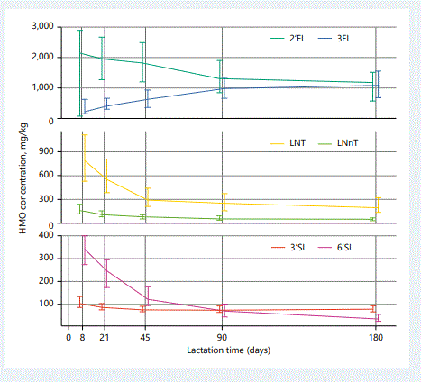
The total HMO content in milk decreases during lactation, presumably reflecting the changing needs of the developing infant. HMO content in colostrum may be over 20 g/L, decreasing to around 13 g/L at 4–6 months of lactation [2]. Recent research suggests that the HMO content may slightly increase again during the second year of lactation [3]. The behaviour of individual HMO are not the same (Fig. 1). For example, while the concentration of 2’fucosyllactose (2’FL) decreases over the first few months of lactation, the concentration of 3FL increases. Also those that decrease do not all decrease at the same rate. 6’sialyllactose (6’SL) is generally regarded as the predominant SL in human milk. While this is true at early stages of lactation, the concentration of 6’SL reduces much more dramatically than that of 3’SL, leading to an increased proportion of 3’SL at later stages of lactation. The differing rates of syntheses of individual HMO suggests that they most likely play different biological roles, adapting to the growing infant’s physiological needs [4]. As our knowledge of the dynamics and functions of the different HMO during infant development increases, we will better understand the changing needs for these in the growing infant’s diet.

Fig. 1. Median levels with interquartile ranges over time of lactation of key individual HMO, representative for the fucosylated (2’FL, 3FL), neutral non-fucosylated (LNT, LNnT), and sialylated (3’SL, 6’SL) HMO [5]. 2’FL, 2’-fucosyllactose; 3FL, 3-fucosyllactose; LNT, lacto-N-tetraose; LNnT, lacto-N-neotetraose; 3’SL, 3’-sialyllactose; 6’SL, 6’-sialyllactose
References
1. Oliveira DL, Wilbey RA, Grandison AS, Roseiro LB: Milk oligosaccharides: a review. Int J Dairy Technol 2015;68:305–321. http://dx.doi.org/10.1111/14710307.12209.
2. Coppa GV, Gabrielli O, Pierani P, Catassi C, Carlucci A, Giorgi PI: Changes in carbohydrate composition in human milk over 4 months of lactation. Pediatrics 1993;91:637–641.
3. Perrin MT, Fogleman AD, Newburg DS, Allen JC: A longitudinal study of human milk composition in the second year postpartum: implications for human milk banking. Matern Child Nutr 2017;13. http://dx.doi. org/10.1111/mcn.12239.
4. German JB, Freeman SL, Lebrilla CB, Mills DA: Human milk oligosaccharides: evolution, structures and bioselectivity as substrates for intestinal bacteria. Nestle Nutr Workshop Ser Pediatr Program 2008;62:205–218; discussion 218–222. http://dx.doi.org/10.1159/000146322.
5. Austin S, De Castro CA, Benet T, Hou Y, Sun H, Thakkar SK, VinyesPares G, Zhang Y, Wang P: Temporal Change of the Content of 10 Oligosaccharides in the Milk of Chinese Urban Mothers. Nutrients 2016; 8: 346. http://dx.doi.org/10.3390/nu8060346.

