Human Milk Oligosaccharides (HMO): Factors Affecting their Composition and their Physiological Significance

Abstract
Human milk oligosaccharides (HMOs) are elongations of the milk sugar lactose by galactose, N-acetylglucosamine, fucose and sialic acid. The HMOs composition of brestmilk is strongly influenced by polymorphisms of the maternal fucosyltransferases, FUT2 and FUT3, and by stage of lactation. Clinical observational studies with breastfed
infant-mother dyads associate specific HMOs with infant gut microbiota, morbidity, infectious diarrhea and allergies.
Observational and basic research data suggest that HMOs influence the establishment of the early life microbiota and mucosal immunity and inhibit pathogens, thereby contributing to protection from infections. Clinical intervention trials with infant formula supplemented with the single HMO, 2’-fucosyllactose (2’FL) or with 2 HMOs, 2’FL and Lacto-N-neotetraose (LNnT), demonstrated that they allow for age appropriate growth and are well tolerated. A priori defined exploratory outcomes related feeding an infant formula with 2 HMOs to fewer reported lower respiratory tract illnesses and reduced need for antibiotics during the first year of life, compared to feeding a control formula. In parallel, the early life microbiota composition shifted towards that of breastfed infants. Together, HMOs likely contribute to immune protection in part through their effect on the early life gut microbiota, findings that warrant further clinical research to appreciate our understanding of HMO biology and significance for infant nutrition.
Introduction
What are human milk oligosaccharides (HMOs)? What is their importance for infant nutrition? These questions have intrigued scientists and pediatricians alike for over a century. Advances in analytics as well as large-scale synthesis technologies stimulated great progress in recent years. These provided the materials and tools that enabled the detailed and accurate measurement of HMO quality and quantity, and the study of HMOs in basic research models, and through clinical observational studies and intervention trials.
“HMOs are not HMOs”, meaning that one specific HMO is not equal to another HMO, especially when considering structure function relations. Chemically, HMOs are elongations of the milk-specific sugar lactose in different linkages by one or a combination of the following monosaccharides: L-fucose (Fuc), D-galactose (Gal),
N-acetyl-D-glucosamine (GlcNAc) and N-acetylneuraminic acid (sialic acid, SA). Gal and GlcNAc generally elongate lactose as a disaccharide Gal-GlcNAc. The numerous and diverse HMOs produced might be categorized by specific structural features brought about by different glycosyltransferases involved in their synthesis. However, many HMOs combine different structural features.
Breastmilk, the recommended and naturally adapted nutrition for infants, is associated with a reduced risk for infection- related illnesses and possibly for diabetes and overweight, while the situation for allergies is less clear [1]. This suggests that breastmilk-specific components such as HMOs and other bioactives, may contribute to such benefits.
Due to their structural similarity with mucosal glycans and their non-digestible nature, HMOs expectedly affect numerous glycan-mediated processes like the colonization of the early life microbiota and the infectivity of pathogens (Figure 1A). Based on clinical observational and basic research data, HMOs, act in a structure-function specific way helping the (i) establishment of mucous adapted microbiome, (ii) resistance to pathogens and (iii) reactivity of the mucosal barrier and immunity, thereby contributing to immune protection.
Here, we briefly review genetic and environmental factors affecting HMO composition in breastmilk and the physiological role of HMOs as supported by clinical observation studies, preclinical research on mode of action and insights from clinical intervention trials.
Maternal glycosyltransferase polymorphisms affect HMOs composition.
HMOs resemble the blood group antigens and further sialylated glycans that cover the human mucosa. The same glycosyltransferases are generally involved in the synthesis of mucosal cell glycans and mammary gland expressed HMOs. The fucosyltransferases FUT2 (Secretor gene) and FUT3 (Lewis gene) are the best described due to their natural polymorphisms in humans [2] (Figure 2A). Specific genetic polymorphisms abolish their respective enzyme activity. Thus, specific HMOs structures that depend on FUT2 or FUT3 can be identified. FUT2-dependent HMOs all contain alpha-1,2 linked fucose, for example 2’FL, lactodifucosyllactose (LDFT), Lacto-N-fucosylpentose-I (LNFP-I). Interestingly, trace amounts of 2’FL were found in breast milk of presumed FUT2 negative mothers in Asian populations [3, 4], indicating that the nature of these inactivating polymorphisms and thus the HMO profile may be population-specific [5]. Typical FUT3 dependent HMOs are LNFP-II, lacto-N-difucosylhexose-I (LNDFH-I) containing alpha-1,4 linked fucose and to a lesser extent 3-fucosyllactose (3FL) and LDFT containing alpha-1,3 linked fucose. In breastmilk that does not contain any detectable LNFP-II, reduced amounts of HMOs with alpha-1,3 fucose on glucose and increased amounts of those with an alpha-1,3 fucose on GlcNAc are found. Hence, another FUT (e.g. FUT4, 5, 6, 7 or 9) is also involved in the formation of HMOs with an alpha-1,3 linked fucose on GlcNAc and glucose.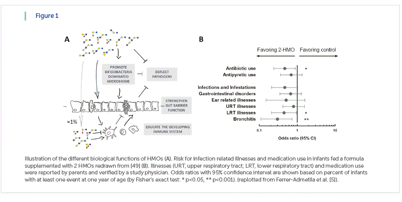
The absence of a functional FUT2 or FUT2 and FUT3 affects the concentration of total HMOs in milk, when expressed as the sum of all quantified HMOs [2] (Figure 2). While some HMOs increase when FUT2 is missing (e.g. LNT, 3FL), in the absence of fucosylation additional larger non-fucosylated HMOs might also be produced.
To date, no common genetic polymorphisms for sialylated HMOs are described, indicating that if inactivating polymorphisms in sialyltransferase genes exist, they are extremely rare. From mouse studies, the sialyltransferases ST6Gal1 and ST3Gal4 are involved in the synthesis of 6’-sialyllactose (6’SL) and 3’-sialyllactose (3’SL) respectively, with a further sialyltransferase, probably ST3Gal1, also making 3’SL [6].
Another mechanism affecting HMO composition is probably the donor and acceptor substrate availability as suggested by the increase of 3FL when the major fucosyl-HMO 2’FL decreases in concentration [3].
Interestingly, HMO concentrations change during stage of lactation with different HMOs showing different dynamics [3]. HMOs like 6’SL or LNT decrease more rapidly during the first weeks of lactation, while 2’FL and 3’SL, for example, decrease more slowly over a longer time period and again others, like 3FL, actually increase in concentration with time of lactation (Figure 2B).
Such compositional changes due to the genetic background of mothers and stage of lactation can confound observations relating HMOs to clinical parameters in the breastfed infants and, therefore, need to be considered.
HMOs composition and infant gestational age, maternal diet and physiological state.
HMOs concentrations in colostrum, transitional and mature milk seem not to change between mothers giving birth to preterm (n=18; gestational age <37 weeks) and term (n=14; gestational age ≥37 weeks) infants [2]. Further, fucosylated and sialylated HMOs were reported to be similar between preterm and term milk, although preterm milk seemed more variable in the expression of fucosylated HMOs [7].
Today, we do not know whether and how maternal diet might influence HMO composition. A recent observational study with 33 breastfeeding mothers and their infants from The Gambia, Africa, reported a significantly higher HMO content in milk at 20 weeks of lactation in the dry season (n=21) compared to the wet season (n=12) [8]. The authors propose a possible link to the higher energy intake during the dry season. In two other African mother-infant cohorts from Malawi (n=88 and n=215), total HMOs and also sialyl-HMOs and fucosyl-HMOs, were lower at 6 months postpartum in breastmilk of mothers having severely stunted infants compared to those with normal size infants [9]. These studies suggest that maternal nutritional and health status may affect HMO composition.
By analogy, higher maternal body mass index and gestational weight gain, generally reflecting an altered metabolic physiology, might affect HMO composition. Studies to this end are currently ongoing [10] (Binia et al. 2017 Abstract at FASEB SRC). Suitable studies are warranted to investigate possible alterations of HMOs composition due to maternal energy and specific nutrient intake.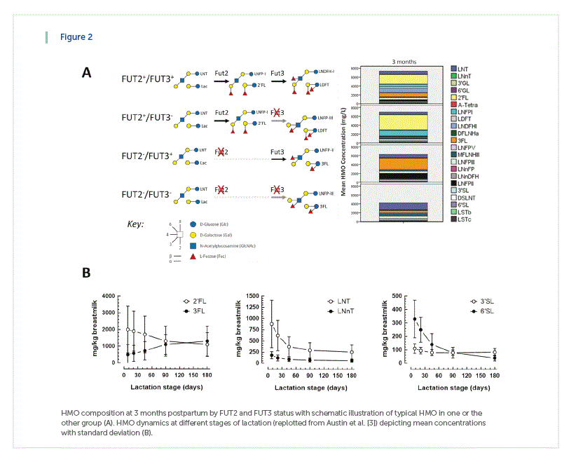
The HMO composition is associated to the establishing gut microbiota in infants.
The early life microbiome has a major impact on the developing immune defenses, itself being an important element by providing pathogen colonization resistance, for example. Interestingly, the establishing intestinal microbiota also contributes, via an innate lymphoid cell-mediated process, to improved protection against respiratory tract infection [11]. The pioneers of human milk and breastfeeding research observed a strong link between breastfeeding and immune protection to infectious morbidity and mortality. Breastfed infants were recognized to harbor an early gut microbiota dominated by bifidobacteria, not seen in formula fed infants, and a human milk specific “bifidofactor” was identified in the HMO fraction of breast milk [12].
From research on early life microbiota, we know that bifidobacteria can utilize and grow on different individual HMOs in a strain-specific way [13, 14]. Several studies observed an increased bacterial metabolic activity upon growth on HMOs, exemplified by the formation of the short chain fatty acid acetate [15, 16]. Noteworthy, numerous potentially pathogenic bacteria from the Enterobacteriaceae group were shown not to grow on individual HMOs as the sole carbon source [17], while growth of other pathogens, like Streptococcus agalactiae (group B Streptococcus, GBS) was shown to be inhibited by HMOs [18, 19].
Recently, LNnT in breastmilk was associated with Bifidobacterium longum ssp infantis abundance [8]. In bi-associated gnotobiotic mice harboring only one
Bacteroides and one B. longum ssp infantis strain, LNnT lead to bifidobacteria dominance although both bacteria could actually use LNnT in vitro [20]. In gnotobiotic mice humanized with seven human microbes, B. longum ssp infantis also showed higher abundance when these mice were fed 2’FL combined with LNnT as compared to LNT alone (Sprenger N. et al. unpublished observation), although B. longum ssp infantis is able to grow on many different HMOs, including LNT, as substrate [13].
Genomic and glycomic analyses in infants provided further evidence for a role of HMOs in shaping the early infant gut microbiome, revealing associations between individual HMOs and bacterial genera in infant stool [21-23]. A bifidobacteria dominated gut microbiota in breastfed infants (n=105) at 4 months of age was associated to breastmilk containing FUT2- HMOs [24]. The FUT2 status of the infants and its possible confounding effects on the infant microbiota profile were not assessed, despite earlier data proposing the FUT2 status itself can influence the gut microbiota at least in adults [25]. In another cohort, the analysis of a relatively small subgroup of 4 month exclusively breastfed infants (n=14) showed an association of maternal FUT2-positive status with higher bifidobacteria abundance up to 2-3 years of age [26]. However, no statistically significant HMO effects on global bifidobacteria shifts were reported in another recent study of 33 Gambian mothers and infants [8], while the abundance of individual bifidobacteria like B. longum ssp infantis still correlated with LNnT concentrations in breastmilk. These first reports reveal the need for larger observational studies of similar design, including comprehensive breastmilk HMO analysis and infant FUT2 phenotyping to gain a more robust understanding of the link between HMO and infant gut microbiome composition.
Today, clinical observations in conjunction with basic research data suggest that FUT2-HMOs, like 2’FL and LNFP-I , but likely other non-FUT2 dependent HMOs, like LNnT for example, are involved in the establishment of a bifidobacteria dominated early life gut microbiota. In vitro studies help to understand HMO-related microbial metabolic capacities and strain specificities, while animal and human observational studies indicate that the interaction between bacteria and the gut mucosa reflect a more complex picture. Hence, with infant health in mind, it is central to gain a better understanding of HMO effects on the microbiome dynamics in their natural ecosystem through a holistic and ecology inspired approach.
HMO composition is linked to infection risk in infants
HMOs were studied in relation with infectious diarrhea incidence in a cohort of Mexican mothers and infants (n=93) [27, 28]. Higher breastmilk concentrations of α1-2 fucosylated HMOs were associated with lower incidence of all causes of moderate-to-severe diarrhea. The most frequently identified cause of diarrhea in the cohort was Cambylobacter jejuni followed by calicivirus and enteropathogenic Escherichia coli. Specifically, higher concentrations of 2’FL and LNFP-I in breastmilk related with a lower incidence of C. jejuni and calicivirus diarrhea respectively. These observations during the breastfeeding period did not persist in the post- breastfeeding period, indicating a possible transient HMO effect in the protection from infectious diarrhea. This fits their presumed role as antiadhesive antimicrobials. Experimental data from preclinical models also show protective effects of 2’FL from C. jejuni [29] and aggregating invasive E. coli [30]. From these data, 2’FL and other FUT2-HMOs seem to act as soluble ligands blocking C. jejuni from adhering to gut epithelial cells, while the protection from E. coli might rather be due to an anti-inflammatory effect, possibly combined with the modulation of the gut microbiota composition.
Glycans containing α1-2-linked fucose expressed on epithelial cells of FUT2 positive infants could act as receptors for pathogen binding, conferring a risk to specific infectious diseases for this population [31]. Genetic studies have shown that infants and children with a non-functional FUT2 gene have strain-specific protection against norovirus and Rotavirus [32, 33]. For specific rotavirus strains, susceptibility depends on FUT2, but also on FUT3 status [34]. Experimentally, infectivity of some rotavirus strains was reduced by the FUT2 HMO 2’FL, while other viral strains were affected by sialylated HMO, namely 3’SL and 6’SL [35]. Similarly, 2’FL also bound to specificn norovirus strains [36].
Besides interfering with pathogen attachment to the host mucosa, HMOs were recently reported to exert bacterial growth inhibitory activities on pathogenic group B Streptococcus (GBS) [18, 19, 37] a major cause of sepsis in preterm infants. Growth of GBS was specifically inhibited by LNT and LNFP-I, while sialylated HMOs or galactooligosaccharides (GOS) had no effect [19]. Experimental data suggests a putative glycosyltransferase of GBS to be involved [19]. Possibly pointing to a similar mechanism, HMOs from milk of a FUT2 negative mother were shown to have bacteriostatic properties via an alteration of biofilm formation [18]. In an observation study of 183 Gambian infant mother pairs, FUT3 positive mothers were reported to be less likely carriers of GBS, as were their infants at birth [37]. Interestingly, infants of FUT3 positive mothers were also more likely to clear GBS colonization from birth to 2-3 months of age compared to infants of FUT3 negative mothers.
In a pilot study of 49 mother-infant pairs, higher breastmilk concentrations of the FUT3 HMOs LNFP-II at 2 weeks were associated with a lower risk of respiratory and gastrointestinal illnesses at 6 and 12 weeks in infants [38]. This association was no longer significant past the breastfeeding period. Similarly, in a nested case cohort study of 143 HIV exposed uninfected children from Zambia, higher concentrations of fucosylated HMOs in breastmilk at 1-month post-partum related to a lower risk of mortality up to 2 years of age [39]. In another small mother-infant cohort from The Gambia (n=33), higher relative breastmilk concentrations of fucosylated HMOs (sum of LNFP-I and LNFP-III) and concomitant lower relative abundance of LNT was associated with lower risk of sickness up to 4 months of age [8].
For respiratory pathogens, direct exposure to HMO would appear less evident and thus any putative HMO-related protection may be mediated by the intestinal microbiome [11, 40]. Yet, experimentally, direct exposure of Streptococcus pneumoniae to LNnT and sialyl-LNnT and subsequent infection effectively blocked its colonization in the lung of a rabbit model [41]. In a cell based assay, LNnT and 2’FL dose dependently reduced Influenza and Respiratory Syncytial Virus (RSV) concentrations within respiratory tract cells [42].
Observational studies together with findings from preclinical models have provided first evidence for an association between HMOs and the risk of infections, mostly in a structure function specific way. Mechanistically, HMOs may act through multiple functions, although preclinical models highlight specific individual functions. The current studies also provide directions to be considered in future observational studies such as timing of milk sampling and breastmilk intake, etiology of infections, quantitative versus categorical analysis of HMOs and finally mother and
infant genetics.
HMO composition might be linked to allergy in infants
Numerous environmental, including nutrition, and genetic factors affect allergies. Among them are breastmilk bioactives, and possibly HMOs. In a cohort of 266 Finnish mother infant pairs with a hereditary allergy risk, 2’FL concentrations in early breastmilk associated with a lower risk to manifest IgE-associated eczema at 2 years of age only in C-section born infants [43]. This observation suggests that 2’FL may influence IgE-associated eczema through the modulation of the early life gut microbiota, known to be different in C-section born infants compared to vaginal born infants. A possible relation of HMOs with cow milk allergy (CMA) was studied in another cohort of 39 mothers with infants who developed CMA by 18 months of age and 41 mothers with infants without CMA [44]. An association was seen between the milk concentration of several individual HMOs (LNFP-III, 6’SL, LNFP-I, DSLNT) and HMO clusters with reduced risk of CMA, with LNFP-III providing the strongest association. Breastmilk sampling varied over the first 6 months after birth and therefore might have introduced a bias, because HMO concentrations change dramatically during this period. Mechanistically, the authors speculate that LNFP-III might act on the immune system via dendritic cells and DC-SIGN. In a preclinical food allergy model, 2’FL and 6’SL were tested and both reduced symptoms involving mast cell activity [45].
The observational studies to date have their limitations, but still provide valuable preliminary data on possible relationships between specific HMOs and risk of allergies. To appreciate such a proposed link requires replication in larger cohorts with harmonized milk sampling, stratification for mode of delivery and evaluation of infant FUT2 and FUT3 genotypes.
Insight from clinical intervention trials with specific HMOs.
Recent progress in industrial biotechnology has made available few individual HMOs, namely 2’FL and LNnT. Preclinical safety toxicity tests established their safety and both obtained approval as novel foods in the European Union and were generally recognized as safe in the USA.
In adults, both 2’FL and LNnT were studied alone or in combination at different doses from 5 to 20 g/day in a placebo controlled, blinded and randomized trial (n=100). Both HMOs were well tolerated and increased bifidobacteria abundance [46].
In infants, two placebo controlled, blinded and randomized clinical intervention trials showed the growth-safety and tolerance of 2’FL combined with either GOS or fructooligosaccharides (FOS) [47, 48] (Kajizer et al. 2016 FASEB J). Infants fed with an infant formula supplemented with 2’FL (0.2 or 1 g/L) combined with GOS or GOS alone showed similar growth as breastfed infants up to 4 months of age (n=314). In a subgroup of infants, immune markers were measured in plasma at baseline and upon stimulation of blood cells with RSV. Globally the immune profile resembled that of breastfed infants when the infant formula was supplemented with 2’FL at the lower or higher dose [48]. Another randomized controlled infant trial showed that an infant starter formula supplemented with 2 HMOs, 2’FL and LNnT, (n=88) allowed for age appropriate growth of term born infants, and was well tolerated when compared to the same infant formula without HMO (n=87) [49]. Interestingly, secondary exploratory findings showed an association between feeding the 2-HMO infant formula and less reported lower respiratory tract illnesses and medication use (especially antibiotics and antipyretics) during the first year of life and beyond the 6 months feeding period. At 3 months, the global microbiota profile shifted in the 2-HMO formula- fed infants away from the control formula fed infants and towards that observed in breastfed reference infants. This shift was mainly due to increases in Bifidobacterium concomitant with decrease in Escherichia and Peptostreptococcaceae [50]. A significantly higher number of infants who were fed the 2-HMO supplemented formula showed a microbiota community structure typical for breastfed infants compared to control formula fed infants, who had primarily a different microbiota community structure. Interestingly, infants with a microbiota community structure typical for control formula fed infants had a 2 times higher risk to use antibiotics during the first year of life compared to those with a microbiota community typical for breastfed infants [51].
These first clinical intervention trials with specific HMOs demonstrate their growth-safety and digestive tolerance. Additionally, as suggested from basic research and observational data, 2’FL and LNnT might contribute to protection from infection-related illnesses and reduced need for antibiotics, possibly through the modulation of the establishing early life gut microbiota.
Conclusion
HMOs composition is affected most notably by the maternal FUT2 and FUT3 status. This is likely due to an evolutionary selective pressure imposed by pathogens or the microbiome at large. Stage of lactation alters HMO composition possibly indicating different infant needs at different extra uterine developmental stages. However, giving birth to a preterm or term infant, who are at different developmental stages, seems not to affect the HMO composition of breastmilk. Clinical observations corroborated by preclinical data and clinical intervention trials support a role for specific HMO in immune protection, primarily from infection related morbidity and use of antibiotics. Further clinical studies, well-designed observational and especially placebo-controlled interventions, are warranted to further substantiate and grow our understanding of the HMO biology and significance for infant nutrition.
References
1. Victora, C.G., et al., Breastfeeding in the 21st century: epidemiology, mechanisms, and lifelong effect. Lancet, 2016. 387(10017):475-490.
2. Kunz, C., et al., Influence of Gestational Age, Secretor, and Lewis Blood Group Status on the Oligosaccharide Content of Human Milk. J Pediatr Gastroenterol Nutr, 2017. 64(5):789-798.
3. Austin, S., et al., Temporal Change of the Content of 10 Oligosaccharides in the Milk of Chinese Urban Mothers. Nutrients, 2016. 8(346):1-22
4. Sprenger, N., et al., Longitudinal change of selected human milk oligosaccharides and association to infants’ growth, an observatory, single center, longitudinal cohort study. PLoS One, 2017. 12(2): p. e0171814.
5. Ferrer-Admetlla, A., et al., A natural history of FUT2 polymorphism in humans. Mol Biol Evol, 2009. 26(9):1993-2003.
6. Fuhrer, A., et al., Milk sialyllactose influences colitis in mice through selective intestinal bacterial colonization. J. Exp. Med, 2010. 207(13):2843- 2854.
7. De Leoz, M.L., et al., Lacto-N-tetraose, fucosylation, and secretor status are highly variable in human milk oligosaccharides from women delivering preterm. J Proteome Res, 2012. 11(9):4662-72.
8. Davis, J.C., et al., Growth and Morbidity of Gambian Infants are Influenced by Maternal Milk Oligosaccharides and Infant Gut Microbiota. Sci Rep, 2017. 7:40466.
9. Charbonneau, M.R., et al., Sialylated Milk Oligosaccharides Promote Microbiota-Dependent Growth in Models of Infant Undernutrition. Cell, 2016. 164(5):859-71.
10. McGuire, M.K., et al., What’s normal? Oligosaccharide concentrations and profiles in milk produced by healthy women vary geographically. Am J Clin Nutr, 2017. 105(5):1086-1100.
11. Gray, J., et al., Intestinal commensal bacteria mediate lung mucosal immunity and promote resistance of newborn mice to infection. Sci Transl Med, 2017. 9(376):1-13
12. Kunz, C., Historical aspects of human milk oligosaccharides. Adv Nutr, 2012. 3(3):430S-9S.
13. Sela, D.A. and D.A. Mills, Nursing our microbiota: molecular linkages between bifidobacteria and milk oligosaccharides. Trends Microbiol, 2010. 18(7):298-307.
14. Matsuki, T., et al., A key genetic factor for fucosyllactose utilization affects infant gut microbiota development. Nat Commun, 2016. 7:11939-11951.
15. Li, M., et al., Microbial composition and in vitro fermentation patterns of human milk oligosaccharides and prebiotics differ between formula-fed and sow-reared piglets. J. Nutr, 2012. 142(4):681-689.
16. Yu, Z.T., et al., The principal fucosylated oligosaccharides of human milk exhibit prebiotic properties on cultured infant microbiota. Glycobiology, 2013. 23(2):169-177
17. Hoeflinger, J.L., et al., In vitro impact of human milk oligosaccharides on enterobacteriaceae growth. J Agric. Food Chem, 2015. 63(12):3295-3302.
18. Ackerman, D.L., et al., Human Milk Oligosaccharides Exhibit Antimicrobial and Antibiofilm Properties against Group B Streptococcus. ACS Infect Dis, 2017. 3(8):595-605.
19. Lin, A.E., et al., Human milk oligosaccharides inhibit growth of group B Streptococcus. J Biol Chem, 2017. 292(27):11243-11249.
20. Marcobal, A., et al., Bacteroides in the infant gut consume milk oligosaccharides via mucus-utilization pathways. Cell Host. Microbe, 2011. 10(5):507-514.
21. De Leoz, M.L., et al., A quantitative and comprehensive method to analyze human milk oligosaccharide structures in the urine and feces of infants. Anal Bioanal Chem, 2013. 405(12):4089-105.
22. Underwood, M.A., et al., Human milk oligosaccharides in premature infants: absorption, excretion, and influence on the intestinal microbiota. Pediatr Res, 2015. 78(6):670-7.
23. Wang, M., et al., Fecal microbiota composition of breast-fed infants is correlated with human milk oligosaccharides consumed. J Pediatr Gastroenterol Nutr, 2015. 60(6):825-33.
24. Lewis, Z.T., et al., Maternal fucosyltransferase 2 status affects the gut bifidobacterial communities of breastfed infants. Microbiome, 2015. 3:13-34.
25. Wacklin, P., et al., Secretor genotype (FUT2 gene) is strongly associated with the composition of Bifidobacteria in the human intestine. PLoS One, 2011. 6(5):e20113.
26. Smith-Brown, P., et al., Mothers Secretor Status Affects Development of Childrens Microbiota Composition and Function: A Pilot Study. PLoS One, 2016. 11(9):e0161211
27. Morrow, A.L., et al., Human milk oligosaccharide blood group epitopes and innate immune protection against campylobacter and calicivirus diarrhea in breastfed infants. Adv. Exp. Med. Biol, 2004. 554:443-446.
28. Newburg, D.S., et al., Innate protection conferred by fucosylated oligosaccharides of human milk against diarrhea in breastfed infants. Glycobiology, 2004. 14(3):253-263
29. Ruiz-Palacios, G.M., et al., Campylobacter jejuni binds intestinal H(O) antigen (Fuc alpha 1, 2Gal beta 1, 4GlcNAc), and fucosyloligosaccharides of human milk inhibit its binding and infection. J. Biol. Chem, 2003. 278(16):14112-14120.
30. He, Y., et al., The human milk oligosaccharide 2’-fucosyllactose modulates CD14 expression in human enterocytes, thereby attenuating LPS-induced inflammation. Gut, 2016. 65(1):33-46.
31. Le Pendu, J., Histo-blood group antigen and human milk oligosaccharides: genetic polymorphism and risk of infectious diseases. Adv Exp Med Biol, 2004. 554:135-43.
32. Thorven, M., et al., A homozygous nonsense mutation (428G-->A) in the human secretor (FUT2) gene provides resistance to symptomatic norovirus (GGII) infections. J Virol, 2005. 79(24):15351-5.
33. Imbert-Marcille, B.M., et al., A FUT2 gene common polymorphism determines resistance to rotavirus A of the P[8] genotype. J Infect Dis, 2014. 209(8):1227-30
34. Nordgren, J., et al., Both Lewis and secretor status mediate susceptibility to rotavirus infections in a rotavirus genotype-dependent manner. Clin Infect Dis, 2014. 59(11):1567- 1573.
35. Laucirica, D.R., et al., Milk Oligosaccharides Inhibit Human Rotavirus Infectivity in MA104 Cells. J Nutr, 2017. 147(9):1709-1714.
36. Koromyslova, A., et al., Human norovirus inhibition by a human milk oligosaccharide. Virology, 2017. 508:81-89.
37. Andreas, N.J., et al., Role of human milk oligosaccharides in Group B Streptococcus colonisation. Clin Transl Immunology, 2016. 5(8):e99.
38. Stepans, M.B., et al., Early consumption of human milk oligosaccharides is inversely related to subsequent risk of respiratory and enteric disease in infants. Breastfeed Med, 2006. 1(4):207-15.
39. Kuhn, L., et al., Oligosaccharide composition of breast milk influences survival of uninfected children born to HIV-infected mothers in Lusaka, Zambia. J Nutr, 2015. 145(1):66-72.
40. Steed, A.L., et al., The microbial metabolite desaminotyrosine protects from influenza through type I interferon. Science, 2017. 357(6350):498-502.
41. Idanpaan-Heikkila, I., et al., Oligosaccharides interfere with the establishment and progression of experimental pneumococcal pneumonia. J. Infect. Dis, 1997. 176(3):704- 712.
42. Geralyn Duska-McEwen, A.P.S., Teah L. Ruetschilling, Edward G. Barrett, Rachael H. Buck, Human Milk Oligosaccharides Enhance Innate Immunity to Respiratory Syncytial Virus and Influenza in Vitro. Food and Nutrition Sciences, 2014. 5(14):1387-1398.
43. Sprenger, N., et al., FUT2- dependent breast milk oligosaccharides and allergy at 2 and 5 years of age in infants with high hereditary allergy risk. Eur. J Nutr, 2017. 56(3):1293-1301
44. Seppo, A.E., et al., Human milk oligosaccharides and development of cow’s milk allergy in infants. J Allergy Clin Immunol, 2017. 139(2):708-711
45. Castillo-Courtade, L., et al., Attenuation of food allergy symptoms following treatment with human milk oligosaccharides in a mouse model. Allergy, 2015. 70(9):1091-1102.
46. Elison, E., et al., Oral supplementation of healthy adults with 2’-O-fucosyllactose and lacto-N-neotetraose is well tolerated and shifts the intestinal microbiota. Br J Nutr, 2016. 116(8):1356-1368.
47. Marriage, B.J., et al., Infants Fed a Lower Calorie Formula With 2’FL Show Growth and 2’FL Uptake Like Breast-Fed Infants. J Pediatr Gastroenterol. Nutr, 2015. 61(6):649-658.
48. Goehring, K.C., et al., Similar to Those Who Are Breastfed, Infants Fed a Formula Containing 2’-Fucosyllactose Have Lower Inflammatory Cytokines in a Randomized Controlled Trial. J Nutr, 2016. 146(12):2559-2566.
49. Puccio, G., et al., Effects of Infant Formula With Human Milk Oligosaccharides on Growth and Morbidity: A Randomized Multicenter Trial. J Pediatr Gastroenterol Nutr, 2017. 64(4):624-631.
50. Alliet, P., et al. Term Infant Formula Supplemented with Human Milk Oligosaccharides (2’Fucosyllactose and Lacto-N-neotetraose) Shifts Stool Microbiota and Metabolic Signatures Closer to that of Breastfed Infants. J Pediatr Gastroenterol Nutr, 2016. 63(1):S55.
51. Berger B, Grathwohl D, Alliet P, et al: Stool microbiota in term infants fed formula supplemented with synthetic human milk oligosaccharides is associated with reduced likelihood of medication (abstract). WCPGHAN, Montreal, 2016.