Influence on Microbiome and Necrotising Enterocolitis – What is new?
Influence on Microbiome and Necrotising Enterocolitis – What is new?
Tim de Meij
Necrotising enterocolitis (NEC) is an inflammation and ischemia of intestinal mucosa. 10% of neonates < 1500 gram develop NEC and the mortality is still very high: 15–30%. There are many complications, e.g. short bowel syndrome, stenosis and developmental delay.
The cause is considered multifactorial: immature gastrointestinal tract is a risk factor, as is enteral feeding, especially formula feeding and colonization with pathogens. Intestinal hypoxia-ischemia-reperfusion is a risk factor too. The staging system of NEC according to Walsh and Kliegman extends from stage I, “suspected”, to stage IIIb, the most severe phenotype. Usually the diagnosis of this disease is established in an advanced stage. So the- re is a great need for novel diagnostic preclinical biomarkers. Combining different biomarkers gives you high accuracy. NEC biomarkers can be found in different fluids, e.g. in urine, blood and even stool, and it is a crosstalk between these biomarkers. E.g. NEC is associated with a change of microbiota.
Microbiota in NEC
Gut microbiota is a key factor in NEC pathogenesis:
- Without bacteria, no NEC
- Antibiotics are still the cornerstone in therapy Probiotics and breast milk are considered protective
- NEC outbreaks usually occur in clusters
- (Early) microbial differences are described between NEC and controls.
This makes microbiota an interesting potential biomarker.
Studies on microbiota and NEC
The largest study of microbiota on NEC thus far (Warner et al, Lancet 2016) was done in 3 US hospitals and included 46 children with NEC > IIb. It showed that increased Proteobacteria were involved or associated with onset of NEC a few days before NEC developed. However, this microbiome risk profile was only present in children GA < 27 wk and, more importantly, in children who developed NEC after 30 days postnatal. This is a very small selective group; the majority develops NEC 2 or 3 wk after birth.
Another study (Heida et al. CID 2016) describes the differences in the microbiome composition al- ready present in the meconium. They followed these children and also saw some differences in the microbiome composition several days before NEC developed. The microbiome signatures in the meconium and in the stool samples before NEC were different. No uniform microbial NEC signature has been detected thus far, limiting its potential for use as a diagnostic biomarker.
The Dutch experience
In the search for novel biomarkers for NEC and sepsis, an ongoing study performed 9 NICUs in the Netherlands and Belgium since 2014. We collect fecal samples from children GA < 30 wk daily up to day 28. Thus far, we have included 1,500 infants and > 20,000 samples. We look at the microbiota before NEC or sepsis develops and we also look at the fecal VOC profile. Volatile Organic Compounds (VOC) are molecules responsible for the smell. VOC are responsible for the odor of stools and > 90% are produced by the intestinal microbiota.
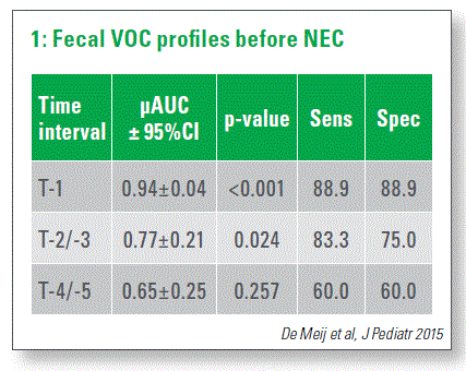
VOC can be detected by various techniques. The most important are chemical-analytical-based techniques such as gas chromatography mass spectrometer, which gives you plenty of information, but is not useful in clinical practice. On the other hand, we have pattern-based techniques such as electronic-nose devices. These are sensors which work similarly to the human nose. We use an electronic-nose device consisting of multiple sensors and based on pattern recognition. It can be used at bedside, so you get direct information within seconds and it is quite cheap. We collect feces samples from NEC, from sepsis patients, and also from controls. In a first pilot study, we compared profiles of 13 NEC patients with 26 controls and we defined different time intervals. When the child diagnosed NEC at day 0, we looked several days back at the VOC and compared it with the sample of children who did not develop NEC. 4–5 days before the diagnosis was made, there was no difference in the profiles. But 2–3 days prior to the onset of NECESSARY, we found increased NEC profiles compared to controls (Chart 1).
The focus of the next study was to compare fecal VOCs from infants who developed late-onset sepsis (LOS). We collected samples from 127 LOS children from these 9 NICUs and we matched them with fecal VOCs from controls. The majority of these infants have CoNS; Staphylococcus aureus was also important.
First of all, we put all these data together and we look to see whether we could discriminate fecal VOCs profiles of children with late-onset sepsis from controls. Clinically, this was not very relevant. We decided to focus on different specific pathogens, so we compared VOCs from gram-negative bacteria and gram-positive bacteria and compared these to the controls. We found that they increased in the days before sepsis.
3 days before Staph aureus sepsis develops, you can see that the VOC patterns of these children are already different from controls and this also accounts for E. coli and Staph.epidermis.
A study by Bos et al. (Plos Pathog. 2013) looked into different VOCs deriving from different pathogens and found that each different pathogen produces only unique VOCs. If you combine all these profiles, then you get rubbish. So that‘s why you have to focus on different pathogens, also in the clinical practice.
In the same group, we looked at the microbiota in the NEC and collected fecal samples from 53 NEC cases and compared them to controls. We performed microbiota analysis up to 3 days prior to the onset of the clinical diagnosis of NEC by means of IS-pro. This technique combines bacterial species differentiation by the length of the 16S-23S rDNA interspace region with instant taxonomic classification by phylum-specific fluorescent labeling of PCR primers (Budding et al FASEB 2010). One of the advantages of IS-pro is that you can get a profile within 5 hours.

Differences in detail
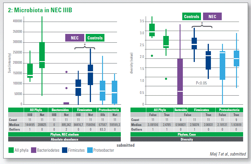
Comparing the NEC profiles in 1, 2, 3 days before NEC to controls was a little bit disappointing be cause we didn‘t see any significant differences at the phylum levels. Then we looked at the diversity of the samples. We found that diversity is also differentiated: first, it is associated with NEC. In the diversity too, we did not see any significant alterations between NEC and controls at 3-1 day prior to NEC at phylum level. Then we looked at the species level, at the cumulative profile of the controls and at NEC: we found no major differences. However, looking into details, you see that the NEC case has more C. perfringens and more S. pyogenes, while controls have a greater abundance of Suterella spp. We took a closer look at the most NEC III phenotype. This outcome is interesting because in this specific group with the most NEC III phenotype, we were able to find differences between NEC and controls which are not present if you look at the overall group. We have a decreased absolute abundance of firmicutes and the difference in the first years as well (Chart 2).
Interestingly, many children among the controls had bacteroides and none of all 55 NEC cases had bacteroides. So it might also play a protective role against the development of NEC. If you look at survivors of NEC for five years, we were able to find microbial differences between NEC and controls.

How could this information be used in the future NICU?
If used to complete NICU population, it might be possible to use one single test with a very high sensitivity and relatively low specificity to select those children who have a higher risk of developing NEC. And if you know which group might develop NEC, you can add another biomarker with a fairly high specificity, e.g. with microbiota: this is more expensive and more complex, but you already need a selection of children. It might be useful for progressively more definite classification. This is just a little bit of dreaming about the future of the NICU, but this is how it might be.
Summary and future perspectives
- It is unlikely that a single biomarker could serve as a standalone diagnostic test for NEC. You probably need a combination of biomarkers.
- Novel predictive biomarkers using proteomic, metabolomics and genomic technologies were evaluated.
- Future diagnostic algorithms based on combination of multiple biomarkers and clinical information
- Machine learning may result in a personalized approach to risk stratification for NEC and discrimination from sepsis.